Recent news
Kaneka Eurogentec completes commercial mRNA..
Company
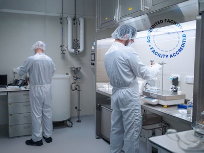
Newly accredited GMP facility for commercial manufacturing of mRNA in anticipation of customer BLA submission....
Cleaner microbial qPCR results start here


Performant dyes for qPCR
and dPCR applications
Up to kg scale plasmids & proteins
for clinical trials and commercialization.
With over 40 years of expertise, we support Dx and testing professionals at every stage.
Company

Products & Services

Exhibitions

11-12 June 2026

22-25 June 2026
We produce plasmid DNA, recombinant proteins and Ab fragments up to kg scale for clinical and commercial human uses.
batch
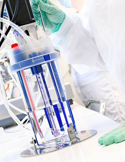
We produce large scale mRNA in our GMP facilities.
We optimized our IVT process to synthesize and purify 50g of material in one batch.
This quantity is equivalent to 100,000’s to millions of doses of mRNA, potentially sufficient for late clinical and commercial uses.
Learn moreWe are pleased to assist you!

Contact our Customer Care, Sales & Scientific Assistance

Consult the most commonly asked questions about our products & services
Browse FAQs
TDS, SDS, guides, posters, brochures, ...
Get assistanceQuality management is fundamental for Eurogentec and is implemented by all personnel in their daily activities.